Plankton life

We often say that the movement is life. For plankton organisms it is really true. Plankton are any organisms that live in the water column and cannot swim against a current. They have to control ciliary activity to stay at a certain depth.Plankton_collage

Depth is so important for the plankton organisms because several environmental factors (temperature, light intensity, the magnitude of UV damage, phytoplankton abundance)  change with depth. Swimming depth influences the speed of organisms’ development and success of feeding . Ciliary swimming is used by the larval stage of many marine invertebrates with a benthic adult. Such ciliated larvae are widespread in the animal kingdom. Among the bilaterians, the lophotrochozoans (e.g., annelids and molluscs) and the deuterostomes (e.g., echinoderms and hemichordates) often have ciliated larvae, while the ecdysozoans (e.g., insects and nematodes) lack them. Outside the bilaterians, cilia-based locomotion is present in ctenophores and in the larval stages of many cnidarians.  Sponges, the basal-most animal group relative to the eumetazoans, often have ciliated larvae. Freely swimming ciliated larvae often spend days to months as part of the zooplankton. The body orientation is maintained either by passive (buoyancy) or active (gravitaxis, phototaxis) mechanisms. When cilia beat, larvae swim upward, and when cilia cease beating, the negatively buoyant larvae sink. The alternation of active upward swimming and passive sinking, together with swimming speed and sinking rate, determine vertical distribution in the water.

Recent investigations of  Markus Conzelmann and his colleagues  from Max Planck Institute for Developmental Biology, Germany have shown that neuropeptides are involved in the regulation of ciliary beating. Neuropeptides have a wide range of functions in the control of neural circuits and physiology, including the modulation of locomotion and rhythmic pattern generation, presynaptic facilitation and remodelling of sensory networks, and the regulation of reproduction. Neuropeptide expression data revealed a cellular resolution molecular map of sensory neurons in the 48 hours after fertilization Platynereis larva. Many neuropeptides also showed intense labelling in the apical neurosecretory plexus of the larva, indicating that they may as well be released there. The innervation of the ciliary band by the diverse neuropeptidergic sensory neurons suggests that these cells, in addition to having a possible neurosecretory function, are involved in the direct motor control of the ciliary band and that there is extensive peptidergic regulation during ciliary swimming.  Experiments show that Platynereis neuropeptides have strong and sequence-specific effects on two parameters of larval ciliary activity: ciliary beat frequency and ciliary arrests. By influencing ciliary beat frequency and ciliary arrests, neuropeptides can change the eventual movement direction, leading to large shifts in the vertical distribution of populations of ciliated larvae.trochophora

Ciliary arrests and beat frequency affect larval swimming. Arrests allow larvae to sink and contribute to the maintenance of an unbiased net vertical displacement. Arrests also lead to irregular swimming tracks. Ciliary beating promotes upward swimming. Neuropeptides can influence both parameters, thereby modulating swimming directionality, the frequency of sinking events, and swimming pattern.

Investigation shows that ciliated larvae use a very simple functional way of regulating swimming by direct sensory-motor innervation.

Such sensory-motor neurons are common in cnidarians. In bilaterians they have been described only in ciliated larvae. This observation raises the interesting possibility that ciliary locomotor circuits in bilaterian larvae have retained an ancestral state of nervous system organization. If this assumption is true, ciliated larval circuitry may give insights into the evolutionary origin of the first nervous systems. The cilia regulatory neuropeptides studied by Markus Conzelmann are widely conserved among marine invertebrates, including other annelids, mollusks, and cnidarians. Because all of these groups have ciliated larvae, these results have broad implications for understanding ciliary locomotor control in a wide range of invertebrate larvae.

M. Conzelmanna, S-L. Offenburgera, A. Asadulinaa, T. Kellera, T. A. Münchb, and G. Jékelya. Neuropeptides regulate swimming depth of Platynereis larvae. PNAS, 2011, vol. 108 no. 46, E1174-E1183

Report by Lena Belikova

What is encrypted in cryptobiosis?

Tardigrades are small animals, about 0,1-1,2 mm in size. They can live in extreme places like deep sea and highest mountains. Tardigrades can survive in very unfavorable environmental conditions. They can stand extreme temperature, pressure, ionizing radiation by entering the unusual stage – cryptobiosis. This amazing ability to tolerate extreme conditions makes tardigrades interesting objects for space, medical, biotechnological research. “Classical” biochemical analytic methods always require destruction of an organism. This investigation shows that SERS spectroscopy opens new way of studying molecular structures on living animals.

In biological research spectroscopy methods are rapidly gaining attention. Raman spectroscopy  is a method based on the inelastic scattering of photons. Photons are good non-invasive instruments to investigate biological objects and processes. Raman spectroscopy delivers vibrational information due to inelastic scattering of photons on the vibrational quantum states of matter. Raman spectra allow to identify proteins, lipids, nucleotides, amino acids, saccharides and shows local distribution of molecules.

Researchers placed plasmonic gold or silver nanoparticles on the surface of objects to prepare them for SERS spectroscopy. For this investigation two large cryptobiotic tardigrades were used. They belong to group of xerophilic tardigrades. This group is inhabiting very unprotected and rapidly drying substrates.  SERS spectra from different species shows significant difference. Spectra from normal and cryptobiosis stages were also different. Investigators registrated changes in protein content and allocation, lipids and nucleic acid bases. And also, Raman signal were registrated in frequency range wich can be related to trehalose. Trehalose is a saccharid presumably a key molecule in cryptobiosis.

Original text Kneipp et al, Surface enhanced Raman scattering on Tardigrada – towards monitoring and imaging molecular structures in live cryptobiotic organisms, Journal of Biophotonics, Volume 6, Issue 10, pages 759–764, October 2013

Report by Elena Belikova

Why fishes don’t like mucus?

Vermetids, or wormsnails, are a small and weakly studied  group of marine gastropods. These molluscs live motionlessly in tube-shaped shells, fixed in substrate. They feed with the help of wide mucus nets catching various floating organisms. This way of feeding is not very unusual in coral reef communities, where vermetids prefer to live.  Different creatures (e.g. other molluscs, some echinoderms and so on) use such nets for passive hunting. Mucus contains a lot of of nutrients, so it’s not surprising that animals eat these nets, especially if they are full  of plankton. Life of some fishes is connected to mucus nets and their producers. In fact, it is very strange that fishes never touch nets of vermetids, even with a lot of food in them. Besides, near the wormsnails’ shells and under their nets corals grow more slowly and sometimes die. These  curious phenomena interested Anne Klӧppel and her colleagues from  University of Stuttgart.

Image

Searching for bioactive substances has become a significant field of biochemistry, molecular biology, microbiology and invertebrate zoology.  Invertebrates, and first of all molluscs, produce a great deal of such matter, for example poison, enzymes, feromones and so on. No wonder that scientists surmised mucus nets contain some substances, looking tastless for fish and decelerating corals growth. But, they weren’t sure,  whether this compounds made by wormsnails  themselves or by  caughted protists, bacteria or algae like dinoflagellate, which are known to be poisonous. To test these hypotheses investigators made experiments with Dendropoma maxima in their natural habitat  in Red Sea and in aquariums. With the help of  high-performance thin-layer chromatography four proteins were isolared. Plankton contamination was excluded by comparing full nets with empty nets.

Image

To check which mucus compounds really have expected effect scientists employed luminescent bacterium Vibrio fischeri. They added each of proteins in plates with cultures of bacterium and observed their lightning. In some plates value  of lightning didn’t change, but in two cases it was inhibited by mucus protein. Thereby predicted bioactive substances were found.

It is the first time, when  bioactive compounds were found within this group. Of course, structure and precise effect of wormsnail mucus should be studied in more detail in future.

Detection of Bioactive Compounds in the Mucus Nets of Dendropoma maxima, Sowerby 1825 (Prosobranch Gastropod Vermetidae, Mollusca), Anne Klöppel, Franz Brümmer, Denise Schwabe, and Gertrud Morlock, Journal of Marine Biology, Volume 2013, Article ID 283506, 9 pages, http://dx.doi.org/10.1155/2013/283506

Report by Khabibulina Valeriya

MARINE GENOMICS: Tell me what’s your larva and I’ll tell you who you are.

The role of plankton in the marine ecosystems worldwide cannot be overestimated. Representing the bottom level of marine food-chains, it provides a crucial source of food for all other marine organisms and influences all universal biochemical cycles including carbon, nitrogen and oxygen cycles. Thus, the high importance of plankton studies becomes evident. Yet it is very difficult to investigate these microscopic organisms. Some of them are algae, others are bacteria, but there is also a large number of larvae in marine plankton. The taxonomic identification of such larvae is a critical step in describing diversity and understanding an ecosystem, but it also is a really difficult task.

net1

Dorothea Heimeier, Shane Lavery and Mary A. Sewell were not afraid of difficulties when they began the studies of Antarctic invertebrate larvae. The goal of the scientific group from New Zealand was to develop and implement a practical approach of identifying larvae using the newest methods of molecular genetics. They studied a wide range of plankton larvae found in Ross Sea, Antarctica, applying a complex of morphological and genetic methods including DNA barcoding.

They worked with over 2000 larval samples and achieved the identification for 35% of all individual specimens, which is a surprisingly good result in comparison with all previous works. The results of this study help to understand marine communities and are a critical step in describing their diversity. Also it provides a reference DNA database for future identification of marine invertebrates.

D. Heimeier, S. Lavery, M. A. Sewell. Using DNA barcoding and phylogenetics to identify Antarctic invertebrate larvae: Lessons from a large scale study. Marine Genomics, Volume 3, Issues 3–4, 2010, Pages 165–177

The report by Yana Frolova

Decolourizing Immunity

Immunity plays a giant role in vertebrates’ interactions with different organisms, both positive and negative. But for studying the evolution of immune system and its emergence we should pay attention to invertebrates, and especially their symbiotic relationships.

Olivier Detournay and his colleagues from Oregon State University have analyzed the participation of TGFβ in different ways of immune-like responses in model anemone Aiptasia pallida. TGFβ, or transforming growth factor beta, is a member of a large superfamily of signaling molecules found throughout the Metazoa that control a lot of cellular functions . Scientists identified homologs to this protein in cnidarians where they have been involved in many processes but, to date, not in immunity. This study has shown the presence of a tolerogenic host innate immune response involving TGFβ in a cnidarian–dinoflagellate mutualism. The goal of this work was to provide evidence of TGFβ pathway components in a symbiotic cnidarian and to explain their role in a tolerogenic immune mechanism that regulates this partnership. Scientists hypothesized that symbionts cause upregulation of a TGFβ. The following modulation of an immune response results in persistence of symbionts in host tissues.

Such a symbiotic system is an important part of marine ecosystems because of coral reefs’ formation. This intracellular association is based on nutrient exchange and is essential for both partners to thrive in nutrient-poor tropical seas. Cnidarian hosts, such as corals and anemones, harbor photosynthetic dinoflagellate endosymbionts from the genus Symbiodinium. Although cnidarian–dinoflagellate symbioses are stable in non-stressed conditions, various environmental factors, most notably elevated temperature caused by global warming, can break partnership leading to loss of symbionts from host tissues. This phenomenon, known as coral bleaching, results in greatly reduced host fitness and can lead to reef destruction. The cellular mechanisms causing symbiosis dysfunction and bleaching are largely unknown, but recent studies indicate that reactive oxygen and nitrogen molecules, host innate immunity and host cell apoptosis all play a role.

This investigation proved a TGFβ role in cnidarian–dinoflagellate mutualism. In the experiment anemones without simbionts in presence of anti-TGFβ factors were occupied by simbionts faster then control group in clean water. Moreover, it was shown that NO can cause increase of TGFβ in host-tissue and, therefore, lost of dinoflagellates and bleaching. So, in Cnidaria TGFβ homologs take part in microorganisms’ resistance and tolerance like in humans.

O. Detournay, C. E. Schnitzler, A. Poole, V. M. Weis
Regulation of cnidarian–dinoflagellate mutualisms: Evidence that activation of a host TGFb innate immune pathway promotes tolerance of the symbiont. Developmental and Comparative Immunology, 2012. 38: 525-537

The report by Lera Khabibulina

They look at you with wide-open arms

Echinoderms in general, and especially the brittlestars  (Ophiuroidea), show a wide range of responses to light intensity, from a largely light-indifferent behaviour to evident colour change and rapid escape behaviour. Joanna Aizenberg and Alexei Tkachenko from Bell Laboratories/Lucent Technologies, together with Murray Hill from New Jersey and other collegues from Israel and Los-Angeles, made a research of photosensivity among some Ophiuroidea.

Analysis of the skeletal structure of two Ophiocoma species reveals two extreme photosensitivity types. Ophiocoma pumila shows no colour change and little reaction to illumination. Ophiocoma wendtii is a highly photosensitive species, and it changes colour markedly from homogeneous dark brown during the day to banded grey and black at night .The upper figure demonstrates that light-indifferent species Ophiocoma pumila shows no colour change from day (left part of the picture) to night (right part). The lower one shows how lightsensitive species O. wendtii changes colour markedly from day (left) to night (right).

Another conspicuous behavioural response to light is negative phototaxis: O. wendtii is able to detect shadows and quickly escape from predators into dark crevices. This behaviour is usually associated with the presence of discrete photosensory organs. No specialized eyes have, however, been documented in brittlestars and their reactions to light have been linked to diffuse dermal receptors.

The analysis of arm ossicles in Ophiocoma showed that in light-sensitive species, the periphery of the labyrinthic calcitic skeleton extends into a regular array of spherical microstructures that have a characteristic double-lens design. These structures are absent in light-indifferent species. Skeletal elements of echinoderms are each composed of a single crystal of oriented calcite shaped into a unique, three-dimensional mesh (stereom). In cross-section they have a remarkably regular double-lens shape.

During the research it was estimated that the microlenses are optical elements guiding and focusing the light inside the tissue. The estimated focal distance (4±7mm below the lenses) coincides with the location of nerve bundles of the presumed primary photoreceptors. The lens array is designed to minimize spherical aberration and to detect light from a particular direction. The presence of transparent regions of compact stereom has been reported also for sea stars and sea urchins. The scientists proposed that these calcitic microstructures might have a function in directing and focusing the light on photosensitive tissues. Calcitic microlenses were used by the trilobites .

The demonstrated use of calcite by brittlestars, both as an optical element and as a mechanical support, illustrates the remarkable ability of organisms, through the process of evolution, to optimize one material for several functions, and provides new ideas for the fabrication of `smart’ materials.

J. Aizenberg, A. Tkachenko, S. Weiner, L. Addadi. Calcitic microlenses as part of the photoreceptor system in brittlestars. NATURE, 2001,vol. 412, p.819-822


Report by Yulia Chapurina

How the worm got it’s pharynx

Have you ever walked along a sandy beach on a summer evening? Surely! Chances are, it had never crossed your mind that you were walking on miriads of misterious tiny living creatures. Yet biologists from all over the world find such habitats to be exiting research grounds. Among the meiofauna of sandy and muddy marine sediments and on algae they are searching for small (sometimes microscopic) flatworms – Acoels.

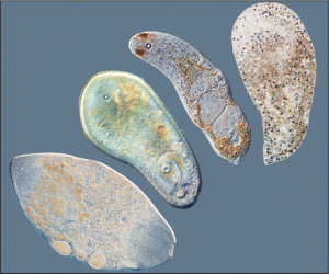
Various acoela worm

These hermaphroditic unsegmented worms only live in marine habitats and are distributed widely around the globe. They constitute their own clade Acoela that includes approximately 380 species. Acoels lack a gut cavity as well as coelomic cavity, excretory and vascular systems. Studying them is quite challenging as the worms are small and rather fragile.

Despite such a simple morphology (acoela have a limited number of organs) they display almost unlimited variability in their configuration. Therefore scientists often consider them as an excellent model group for studies of evolutionary scenarios  and processes.

So do the group of evolutionary morphologists who presented their collaborative paper in Systematic Biology by Oxford University press in August 2011. Inspite of considerable scientific significance, there had been no extensive phylogenetic study of the group till Ulf Jondelius, Andreas Wallberg, Matthew Hooge and Olga Raikova presented their work. They identified morphological traits and molecular biological markers of a large number of species, and also gathered all existing data on several other previously studied species, thus covering 1/3 of the known acoel species. Comparative statistical analysis of such an impressive dataset allowed them to adjust the position of the group on the global phylogenetic tree, to clarify many intraspecific relationships and also allowed to propose the ancestral traits of the whole group.

Authors use 2 nuclear ribosomal genes and 1 mitochondrial gene in combination with 37 morphological characters (such as size, shape, pigmentation, presence or absence of algal simbionts, eyes, pharynx, glands and genitals etc) to built the tree. The hypothetical common ancestral features were proposed after building the Acoel root. Authors used Bayesian approach to determine what features are more likely to be primitive. The ancestral acoel was likely a cylindrical worm with a pharynx located in the posterior end of the body,  relatively simple reproductive system lacking female accessory organs, symbiotic algae, pigmantation and eyes are absent.

The understanding of the phylogenetic tree is impossible without an advanced study of all possible groups of creatures. Such studies move us to understand the whole tree of life.

Authors believe that it will be possible to trace the evolution of morphological traits of this exciting worm group, permitting to resolve many basic question in developmental, morphological and evolutionnary biology.

U. Jondelius, A. Wallberg, M. Hooge And O. I. Raikova. How the Worm Got its Pharynx: Phylogeny, Classification and Bayesian Assessment of Character Evolution in Acoela. Syst. Biol, 2011, 60(6), 845–871

Report by Yana Frolova

Not-so-simple Trichoplax

Trichoplax is the only species in Placozoa group and one of the most mysterious and amusing invertebrate animals. It looks like a thin plate about 4 mm in size and consists of two layers of primitive epithelial cells without basal membrane. Between these layers there are a lot of motile cells that can have different functions… but we don’t quite know which ones. It seem like Trichoplax is a very simple creature: it has no color, no constant body shape and, as some suggest, no symmetry. But it’s biology is not as easy to understand as it seems to be.

20080822427590-362057510

Since Tricholax had been discovered and described by F. Schulze in 1883 it has been posing a great deal of questions to scientists. In first years of observations people didn’t know where it was from because it would just suddenly appear in sea aquaria and escape them . Some scientists were sure that Trichoplax is just unknown Cnidaria or Spongia larvae. Nowadays Trichpoplax is found in Pacific Ocean, but we still don’t exactly understand it’s ecology, food and sex behavior, and especially anatomy and morphology features. However, of main interest for biologists is the systematic position of this not-so-simple animal. Is it a first multicallular animal? Or maybe it’s an ancestor of Spongies, or Cnadaria, or bilaterians?

Mansi Srivastava from University of California  with colleagues from different countries made a whole genome sequencing trying to answer these questions. The size of Trichoplax genome is about 98  million  bp  and it contains 11,514 protein coding genes.  Most of them have a detectable similarity to other animals’ genes and 7800 of these genes are common with bilaterian and cnidarians. Analysis of the exon–intron structure of orthologous genes demonstrated a high degree of conservation in Trichoplax relative to other eumetazoans. Using nine whole-genome sequences of other Metazoan, scientists showed that Placozoan is a sister group to all of them. So demosponge sequences diverge before the Trichoplax–cnidarian–bilaterian clade. But there is no evidence to prove that Trichoplax is either a derived or basal cnidarian or bilaterian.

Moreover, it has been shown that Trichoplax with its simple body and without normal epithelial layer in histological meaning has a lot of genes that encode complex processes appropriate to eumetazoans. For example, there are some transcription factors and signal pathways involving proteins associated with development and cell-type specification.

This work has just been started, but it discovers different possibilities to understand life of Trichoplax and some evolutionary and developmental questions.

M. Srivastava. The Trichoplax genome and the nature of Placozoans. Nature, 2008, vol. 07191. P. 955-961. doi:10.1038

Report by Lera Khabibulina